
The Challenge
The traditional process of analysing cardiac MRI scans is:
- Time-consuming: A cardiac MRI scan (study) has between 3500 and 5000 slices, and manual analysis often takes up to 60 minutes per scan.
- Subjective & inconsistent: Accuracy varies based on radiologist experience and fatigue, often resulting in diagnostic errors or delays.
- Resource-intensive: Many centres lack specialised personnel or infrastructure to process cardiac MRIs effectively.
- Disconnected: Existing solutions are vendor-specific, lack interoperability, and are often incompatible across platforms.
These limitations contribute to decreased accuracy, delays in reporting, and ultimately suboptimal output.
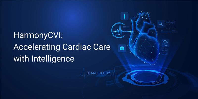
Our Solution
HarmonyCVI is an AI-powered cardiac MRI post-processing platform that automates and accelerates diagnostic interpretation using deep learning.
- Fully automated segmentation and quantification of cardiac anatomy, including volumetric and Q-flow analysis, with clinical-grade accuracy.
- Empowers clinicians with real-time, vendor-neutral, device-agnostic access to insights, reports, and structured data anytime, anywhere.
Key Features
-
- Volumetric Analysis: Automated AI-based contour detection for left and right ventricles (EDV, ESV, etc.)
- Q‑Flow Analysis: Automated segmentation and blood flow/velocity calculation without manual contouring
- Multi‑View Layouts: Customizable composite layouts for syncing functional views for enhanced interpretation
- Advanced Imaging: Supports Delayed Enhancement, Myocardial mapping (T1, T2 & T2*), Extracellular Volume Calculation (ECV), Global Longitudinal Strain, Atrial Volumes Analysis and bookmark-sharing across sessions
Integrated Reporting: Real-time, interactive, customisable reports aligned to clinician needs.
The Impact
- Clinical Outcomes
- <2-minute for processing all the slices and giving contours and parameters, making a quicker turnaround for the radiologist to report compared to traditional 60+ minutes from a manual process to about a 10-minute turnaround
- Reduced radiologist reporting time from 60+ minutes to ~10 minutes, processing all slices, contours, and parameters in under 2 minutes, streamlining workflow and boosting efficiency
- Accurate analysis for cardiac MRI workflows
- Enables faster triage and better therapeutic decisions
- Clinician Efficiency
- Seamless integration into clinical workflows
- Reduces radiologist burden and minimizes inter-reader variability
- Supports reimbursable, billable diagnostic workflows
- Health System Benefits
- Adaptable for large hospitals, private clinics, and remote settings
- Reduces time-to-treatment, lowers risk of missed diagnoses
Technologies Used
- Front-End Technologies
- AngularJS: A component-based web framework for dynamic and responsive UI.
- Weasis Viewer: Embedded DICOM viewer for rich, interactive medical image visualisation directly in the browser.
- Back-End Technologies
- Java: Core service architecture, handling application logic, orchestration, and system integration.
- AI: Multiple pre-trained deep learning and transformer models like SAM3, MedSAM2, Streamlined MLOps with CubeFlow.
- Databases & Imaging Infrastructure
- PostgreSQL: Relational database used for secure storage of clinical metadata, user data, and analysis results.
- Redis: In-memory data store for real-time caching and task queuing, enabling low-latency responses.
- PACS: To retrieve and store DICOM files, enabling seamless integration into clinical imaging workflows.
- Cloud Infrastructure & Services (AWS)
- Amazon S3: Secure, scalable storage for all incoming and processed imaging studies.
- AWS Lambda: Manages the lifecycle of the AI inference server, auto-starting/stopping for efficient resource usage.
- Amazon CloudWatch: Real-time logging and system monitoring for platform health and diagnostics.
- EC2 Instances:
- AI Server: Dedicated GPU-enabled EC2 instance for executing AI inference tasks.
- Backend Server: Handles API requests, authentication, data processing, and user management
- On-premise: Entire platform is installed on Mac Mini and a few other GPU-based Edge devices
- GPU-enabled machine
- AI Server: Docker instance for executing AI inference tasks
- Backend Server: A Docker instance handles API requests, authentication, data processing and user management and connects to the client’s PACS server.
- GPU-enabled machine
Conclusion
HarmonyCVI redefines how cardiovascular imaging is delivered. By combining AI & deep learning, cloud-native infrastructure, and clinician-first design, it enables rapid, accurate, and accessible cardiac MRI interpretation. With scan-to-report times under 2 minutes and diagnostic accuracy above 99%, HarmonyCVI transforms the cardiac imaging workflow from a manual bottleneck into a scalable, intelligent diagnostic tool.
It’s more than automation; it’s intelligence, delivered.